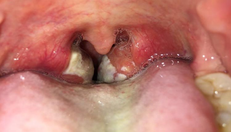
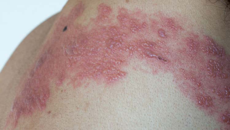
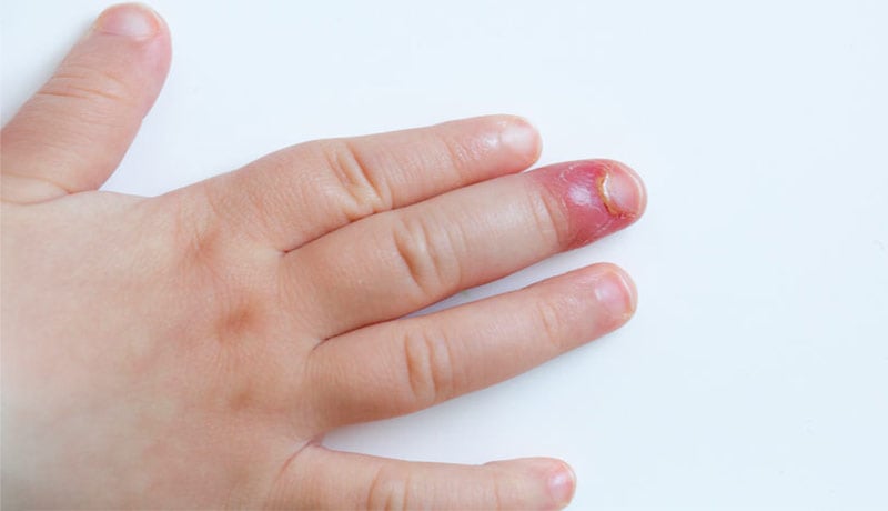
(!) This is a photo page with typical syphilis lesions in their different stages. For detailed information on causes, symptoms and treatment options, visit the article: Syphilis: Symptoms, Tests, Transmission, Treatment, and Cure.
What is syphilis?
Syphilis is a sexually transmitted disease caused by the bacterium Treponema pallidum.
The hallmark symptom of syphilis, typically seen in the initial stage, is a painless ulcer, known as a chancre, predominantly appearing in the genital region. The most significant risk of transmission comes from individuals in the primary or secondary stages of the disease, particularly when active lesions are present on their sexual organs.
Before showing the pictures, here’s a succinct overview of the three distinct stages of syphilis.
Stages of Syphilis
Weeks or months after the hard chancre disappears, syphilis reappears, now in a secondary phase, and can cause lesions on the skin and mucous membranes. If left untreated, the lesions again disappear spontaneously, returning years later in the form of tertiary syphilis, the most serious form of this disease, with a high risk of causing disfiguring lesions.
In the photo gallery below we show images of syphilis in the primary, secondary and tertiary stages. The severe tertiary syphilis lesions shown below are rare today, as most patients receive adequate treatment before the disease reaches such an advanced stage. In the pre-antibiotic era, however, this type of disfiguring lesion was relatively common.
In all three stages of syphilis, treatment is indicated with penicillin.
Caution: The following gallery contains graphic images depicting the primary, secondary, and tertiary stages of syphilis. These images consist of visually intense and disfiguring lesions or representations of sexual organs. We encourage discretion and recommend that you refrain from accessing this gallery if your current environment is not conducive to the viewing of such content.
Images and photos
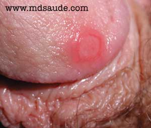
Primary syphilis – Syphilitic chancre
The primary stage of syphilis is the best known and is characterized by the presence of a genital ulcer called a hard chancre. In the case of transmission through oral sex, the hard chancre can appear in the mouth or tongue.
The initial lesion of primary syphilis manifests itself as a small elevation on the skin of the genitals, which in a few hours turns into an ulcer, usually not painful.
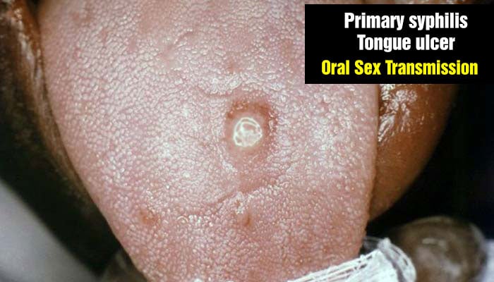
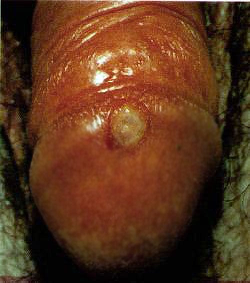
In women, this lesion can go unnoticed due to its small size (on average 1 cm in diameter), the absence of pain and its discreet location between the pubic hair or inside the vagina.
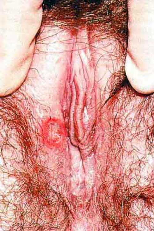
After contamination, it takes an average of 2 to 3 weeks for hard cancer to appear. However, there are cases where this interval can be as short as three days or as long as three months.
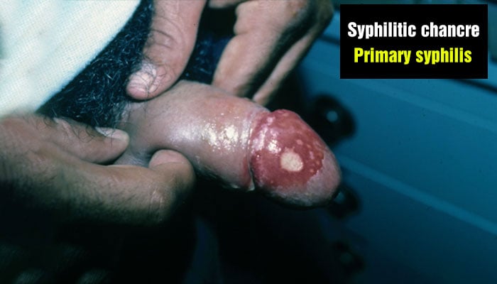
Hard chancre is a lesion that lasts 3 to 6 weeks and disappears even without treatment, which leads to the false impression of a spontaneous cure of the disease.
Secondary syphilis
In 25% of patients not treated in the primary phase, syphilis returns after months or years in silence in the form of secondary syphilis.
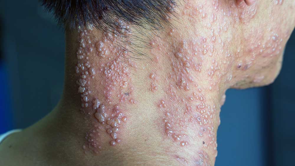
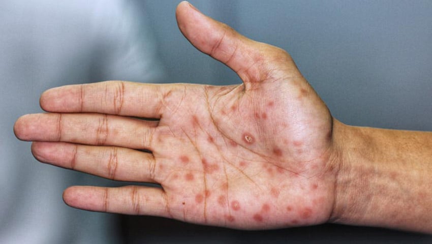
Secondary syphilis typically manifests as a rash on the skin, classically affecting the palms of the hands and soles of the feet. Other common symptoms include fever, malaise, loss of appetite, joint pain, hair loss, eye lesions, and generalized lymph node enlargement.
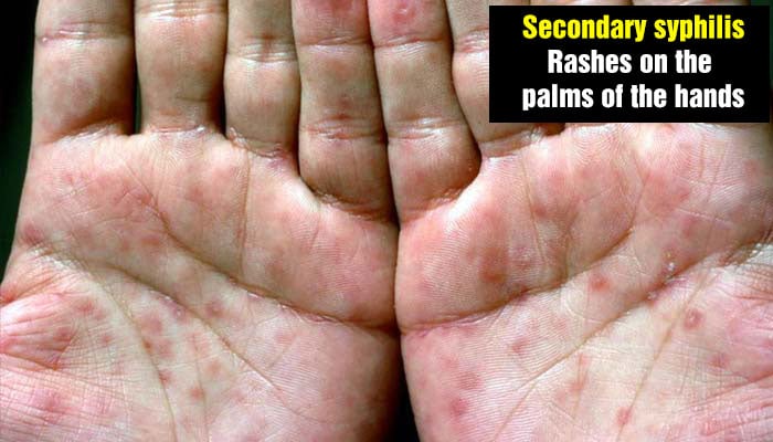

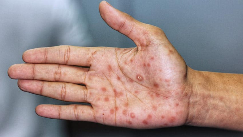
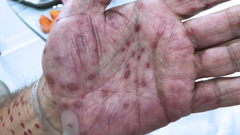
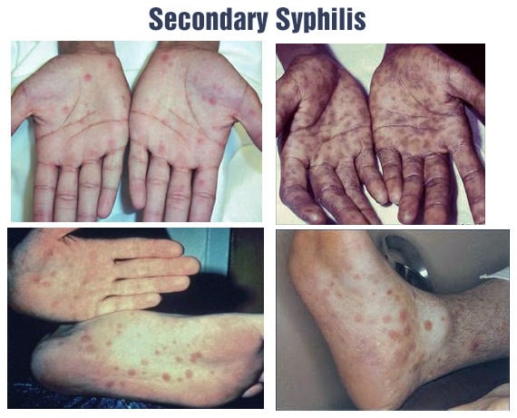

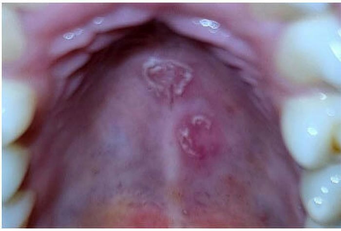
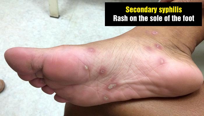
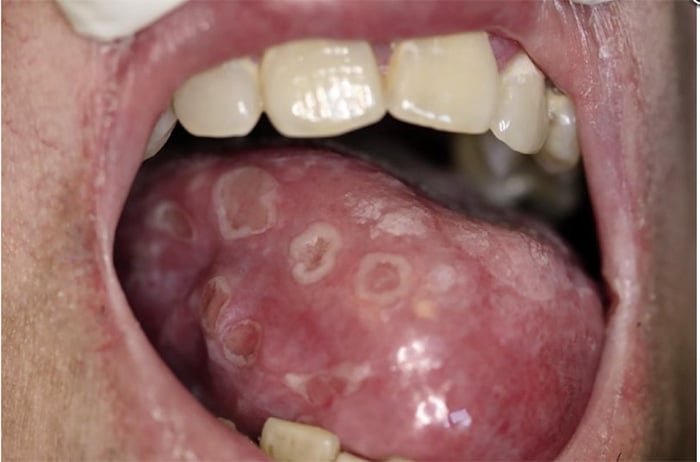
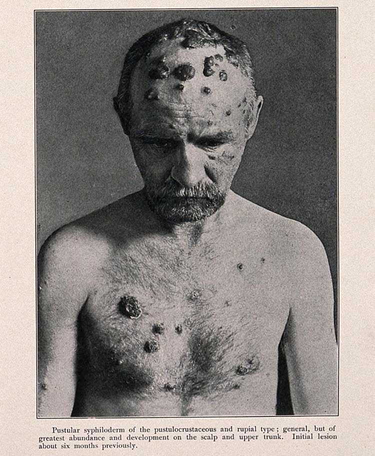
Even without treatment, lesions from the secondary phase may disappear spontaneously. When this occurs, the patient enters the latent phase of the disease. Although there are no symptoms during this phase, laboratory tests for syphilis remain positive.
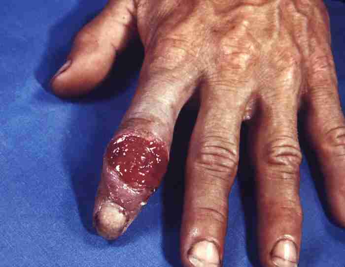
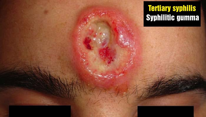
Tertiary syphilis – Syphilitic Gumma
Patients can remain asymptomatic for several years, even decades, during the latent phase before the disease reemerges. This new stage, when symptoms return, is known as tertiary syphilis, the most severe form of the disease.
The tertiary phase presents with three main types of manifestations:
- Syphilitic gummas: large ulcerative lesions that can affect the skin, bones, and internal organs.
- Cardiovascular syphilis: involvement of the aorta, leading to aneurysms and damage to the aortic valve.
- Neurosyphilis: affects the nervous system, causing dementia, meningitis, strokes, and motor problems due to spinal cord and nerve damage.



Credits
- Shutterstock.com
- Multiple skin ulcers from malignant syphilis – The Lancet
- Secondary Syphilis in Cali, Colombia: New Concepts in Disease Pathogenesis – Scientific Figure on ResearchGate. Available from: https://www.researchgate.net/figure/Palmar-and-plantar-rash-of-secondary-syphilis-Typical-palmar-and-plantar-rash-of_fig8_44630365 [accessed 26 Apr, 2023]
- Morais, Lima & Melo, Thayná & Kitakawa, Dárcio & Da, Felipe & Peralta, Felipe & Gonzales, Sabrina & Carvalho, Luis Felipe & Carvalho, Silva. (2022). Secondary syphilis in oral cavity: Case report and literature review. International Journal of Case Reports and Images. 13. 226-229. 10.5348/101366Z01TM2022CR.
- Tertiary syphilitic ulceration of the scalp – St Bartholomew’s Hospital Archives & Museum.
- An Alaskan Inuit skull, showing the effects of syphilis. Photograph by Ales Hrdlicka, ca. 1910.
- A man suffering from syphilis, displaying pustular syphiloderm lesions on his scalp and torso. Process print after a photograph, ca. 1905.
- Face of a woman with a typical ‘syphilitic nose’. Picture: St Bartholomew’s Hospital Archives & Museum.
Author(s)
Médico graduado pela Universidade Federal do Rio de Janeiro (UFRJ), com títulos de especialista em Medicina Interna e Nefrologia pela Universidade Estadual do Rio de Janeiro (UERJ), Sociedade Brasileira de Nefrologia (SBN), Universidade do Porto e pelo Colégio de Especialidade de Nefrologia de Portugal.


